X-Ray
What is radiography?
Radiography is one of the medical imaging methods that can be used to see the inside of the human body in a way and thus help in the correct diagnosis of the disease.
This imaging is done by x-ray radiation. X-rays pass through the skin and hit the sensitive screen. The color of this part can be seen black after the appearance.
On the contrary, it blocks the passage of X-rays and those areas can be seen white after appearing. For this reason, in a radiograph, bones that are denser and stronger are whiter and others are less white.
Areas such as the air or the inside of the intestine where X-rays pass completely without any problems are completely black, and others that slightly block the passage of the rays are seen as gray.
Plain radiography
Plain radiography is the first type of imaging, where the image is recorded as a black and white image on a photo or radiology film, which is a transparent plastic sheet.
The doctor puts this sheet in front of the light and looks at the image. Plain radiography is a very valuable tool for examining bones, their fractures and various diseases.

Digital radiography
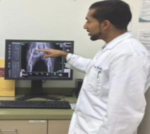
In this type of radiography, the prepared image is sent to the computer and the doctor sees the image on the computer monitor screen. Of course, the images can be printed on the film.
The difference between this method and the old method is, the difference in the radiographer’s film. The same difference between new digital cameras and old cameras.
In digital imaging, instead of plastic film, an electronic screen with millions of small semiconductors sensitive to X-rays is used. X-ray radiation to semiconductors creates an electric charge. Each of the different points of the screen or its pixels receives a specific and different amount of X-rays, and according
to the amount of radiation, the electric charge produced is also different. It is collected many times and sent to the computer so that the pixel information is put together and the image is formed.

